Risk and Control- Melanocyte
Medical cosmetic treatment involves medical assessment because procedures that heat, remove or deliberately injure selected skin structures can also affect healing and pigmentation. Understanding the normal function of melanocytes helps explain why a treated area may become darker or lighter afterwards. Pigment change is a possible complication rather than an inevitable response, and the risk varies between individuals and procedures.
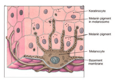
Melanocytes are specialised cells found mainly in the basal layer of the epidermis. They produce melanin inside structures called melanosomes and transfer melanin-containing granules through dendritic processes to nearby keratinocytes. Melanin contributes to skin, hair and eye colour and absorbs some ultraviolet radiation, helping to protect cellular DNA. It does not replace external sun protection. Differences in skin colour mainly reflect the amount and type of melanin produced and how melanosomes are distributed, rather than simply whether melanocytes are present.
Lasers, light devices, chemical or fruit-acid peels, microneedling, radiofrequency procedures and other treatments may produce controlled heat or injury. The associated inflammatory response can stimulate melanocytes and increase melanin production or transfer, leaving the treated area darker. This is known as post-inflammatory hyperpigmentation (PIH). If inflammation disrupts the junction between the epidermis and dermis, pigment may also be deposited more deeply and can take longer to fade. These pathways are complex, and the course cannot be predicted precisely for every person.
Melasma also requires particular care. It is a chronic, trigger-sensitive pigmentary condition that may be aggravated by ultraviolet exposure, visible light, heat, inflammation or some procedures. Lasers and light-based treatments may be considered selectively, but they are not a guaranteed solution and can sometimes worsen pigmentation. The diagnosis, current activity of the condition and treatment options should therefore be assessed individually.
Hypopigmentation, sometimes described as “lightening” or “white spots”, can occur when melanin is temporarily reduced, melanocyte activity is suppressed, melanocytes are injured, or more extensive tissue damage affects normal pigment production. Repeated or unsuitable exposure to a pigment-targeting laser, including Q-switched 1064 nm treatment, has been associated with mottled hypopigmentation. Risk is not determined by energy alone; wavelength, fluence, spot size, treatment interval, cumulative exposure, skin condition and technique may all be relevant. Some areas gradually repigment, while others persist and may be difficult to treat. If deeper injury causes scarring, the structural change may be lasting, although the appearance of some scars can be improved.
People with melanin-rich skin, often represented by higher Fitzpatrick phototypes, tend to experience PIH more frequently, but skin type alone does not determine suitability. Recent tanning or sunburn, melasma or another pigmentary disorder, a previous pigment response, active inflammation, infection, open wounds, abnormal scarring, impaired healing, medication use and the planned depth or type of procedure may all affect risk. Lighter skin can also develop hyperpigmentation, hypopigmentation, burns or scars. Radiofrequency does not target melanin in the same way as a pigment-selective laser, but heat or needle injury can still cause inflammation and pigment change.
Before treatment, a doctor should assess the skin concern, skin type, recent ultraviolet exposure, medical and medication history, previous reactions and the proposed device or procedure. Sun avoidance and broad-spectrum photoprotection before and after treatment may reduce pigment-related risk, but they cannot eliminate it. Prescription retinoids or depigmenting agents are not routinely suitable for everyone and may irritate the skin; their use and timing should be determined according to the diagnosis, procedure, pregnancy status and individual skin condition. After treatment, follow the clinician’s wound-care and sun-protection advice, and seek medical review if there is increasing pain, blistering, discharge, delayed healing or an unexpected change in pigmentation.